Neuroendoport® Clinical Case Study
The Patient
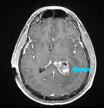
A 47-year-old man was suffering from relentless headaches, confusion, and visual disturbances. An MRI showed a large intraventricular tumor.
|
Pre-surgical scan shows a 2-inch intraventricular tumor.
|
|
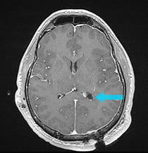
After successful surgery, the ventricle is visible on the scan as a dark space.
|
The Challenge and Solution
The intraventricular tumor was nearly two inches in diameter. Because of its large size, the tumor was removed by two operations on two separate days using the Neuroendoport technique. The pathology report showed a subependymoma, a benign tumor in the ventricle. Surgical removal was achieved with minimal manipulation of the surrounding brain, allowing the patient's speech and vision function to be maximally preserved.
The Result
The post-surgical MRI shows complete removal of the tumor. The patient's confusion and headaches resolved, with just minor visual difficulty remaining.
Treatment and results may not be representative of all similar cases.


