Skip to Content

Mitral Valve Disease

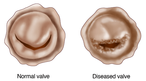
The mitral valve, located between the heart's left upper and lower chambers, has two flaps. These flaps open to allow blood to pass from the atrium to the ventricle. When the heart relaxes and the ventricle fills, the pressure causes the flaps to close.

Mitral valve disease occurs when your mitral valve stops opening and closing properly.

The UPMC Heart and Vascular Institute's Center for Mitral Valve Disease is a leader in diagnosing and treating mitral valve disease, including mitral stenosis and mitral regurgitation.

Contact the UPMC Heart and Vascular Institute

To request an appointment, contact the UPMC Heart and Vascular Institute:


What is Mitral Valve Disease?

The mitral valve is located between the heart's upper left chamber (the left atrium) and the heart's lower left chamber (the left ventricle).

When the heart contracts, the mitral valve's two flaps — or leaflets — open to allow blood to pass from the atrium to the ventricle. When the heart relaxes and the ventricle fills, the pressure causes the flaps to close.

Mitral valve disease is when your mitral valve stops opening and closing properly.

Mitral valve stenosis is a narrowing of the heart's mitral valve, which controls the blood flow through the heart. This prevents your mitral valve from opening properly and reduces blood flow out of the left atrium.

Types of mitral valve disease

  • Mitral stenosis is when the valve becomes restricted or partially blocked. This “blockage” prevents some of the blood from moving out of your atrium and into your ventricle.
  • Mitral regurgitation occurs when your mitral valve no longer closes tightly. This allows blood to travel backward, from the ventricle to the atrium. The most common cause of mitral regurgitation or “leakage” is known as mitral valve prolapse.

Causes of mitral valve disease

Causes of mitral stenosis include:

  • As one ages, calcium can build-up around and on the mitral valve. This is the most common form of mitral stenosis.
  • Rheumatic fever and scarlet fever in childhood may cause the leaflets of the valve to thicken or fuse over prolonged periods of time, preventing it from opening properly. This is the second most common cause of mitral stenosis.
  • Other well-known causes of mitral stenosis are:
    • Prior external radiation to the chest cavity during cancer treatment.
    • Prior known history of congenital, or birth-related, abnormalities.
    • Some rare medications.

Causes of mitral regurgitation include:

  • Mitral valve prolapse is a condition when one is born with excess leaflet tissue, known as a “floppy mitral valve.” As one ages, this makes the valve prone to leak. This is the most common cause of mitral regurgitation.
  • Infection, or endocarditis, occurs secondary to bacteria entering the blood stream and attaching themselves to the mitral valve. This often happens in people with mitral valve prolapse. The most common cause of bacteria entering the bloodstream is from dental or oral sources. Endocarditis is the second most common cause of mitral regurgitation.
  • Other well-known causes of mitral regurgitation are: 
    • Rheumatic fever can also lead to scarring of the leaflets to cause the valve to leak.
    • A heart attack or lack of blood supply to the heart muscle can stretch the left ventricle and the mitral valve, causing it to leak. 

Risks of untreated mitral valve disease

Without treatment, mitral valve stenosis can lead to:

  • Heart failure — a condition in which your heart cannot pump enough blood to meet your body's needs
  • Heart enlargement
  • Atrial fibrillation (A-fib)
  • Blood clots (A-fib increases the risk of developing blood clots)
  • Fluid build-up in the lungs

Without treatment, mitral regurgitation can lead to:

  • Heart failure
  • Heart enlargement
  • Common heart rhythm problems such as A-fib
  • Rare heart rhythm problems such as premature ventricular contractions
  • In very rare situations, mitral regurgitation secondary to severe mitral valve prolapse can result in sudden death

Why choose UPMC's Center for Mitral Valve Disease?

Our extensive experience allows us to provide the latest treatments for mitral valve disease, including:

  • Complex valve reconstruction
  • Minimally invasive mitral valve catheter-based techniques, such as mitral valvuloplasty and MitraClip

Our team-based approach to care includes cardiac surgeons, interventional cardiologists, cardiologists, anesthesiologists, and nurses.

Our team focuses on open communication while working with you, your family, and your personal doctors to provide coordinated care to best meet your needs. 

Learn More About Mitral Valve Disease

UPMC Heart and Vascular Institute

From our Health Library

Mitral Valve Disease Symptoms and Diagnosis

Mitral stenosis symptoms

Symptoms of mitral stenosis include:

  • Shortness of breath
  • Fatigue, worse with activity
  • Coughing — can often be a dry cough
  • Swelling of feet or ankles
  • Heart palpitations, which is the sensation of feeling your own heartbeat
  • Frequent infections of the lungs, such as bronchitis

Mitral regurgitation symptoms

Symptoms of mitral regurgitation include:

  • Shortness of breath
  • Fatigue, worse with activity
  • Coughing — can often be a dry cough
  • Swelling of feet or ankles
  • Heart palpitations, which is the sensation of feeling your own heartbeat
  • Chest pain
  • Anxiety

Diagnosing mitral valve disease

In addition to asking you about your medical history, your doctor at the UPMC Heart and Vascular Institute will perform a physical exam.

Often, mitral valve disease causes murmurs, or abnormal sounds of blood flowing through your heart.

Tests your doctor may use to diagnose mitral valve disease include:

  • Echocardiogram, or a heart ultrasound, uses sound waves to create images of the structures of your heart.
  • Trans-esophageal echocardiogram, or “TEE,” is an outpatient test that uses the same sound technology of a regular echocardiogram but performed with within your swallowing tube or esophagus to get very accurate images of your heart valve, often in 3-dimensions.
  • Electrocardiogram, measures the electrical activity of your heart.
  • Chest x-ray, enables your doctor to see the size and shape of your heart.
  • Cardiac catheterization, is an outpatient test that enables the doctor to see blood flow through the heart and its arteries.
  • Magnetic resonance imaging (MRI) scan, which uses a magnetic field and radio waves to create images of your mitral valve. 

Learn More About Mitral Valve Disease Symptoms and Testing

From our Health Library

Mitral Valve Disease Treatment

Goals of treatment for mitral valve disease are to limit or eliminate the blockage or leakage of your valve while providing symptom relief and improvement of longevity and quality of life.

If the mitral valve disease is not severe, medications and close follow-up with frequent echocardiograms may be possible. However, since mitral valve disease is a mechanical problem with the flow of blood through your heart, it is often best treated with fixing the valve surgically.

Mitral valve repair for mitral regurgitation

The first line treatment of mitral regurgitation, particularly for mitral valve prolapse, is mitral valve repair. This involves surgically restoring the normal function of the mitral valve by rebuilding one’s existing leaflets using several techniques tailored to the individual anatomy.

At UPMC’s nation-leading Center for Mitral Valve Disease, surgeons perform this operation every day with an overall repair rate over 98 percent.

Mitral valve repair can often be performed using minimally invasive approaches through a small incision on the front of the chest, or, in some cases, through a small incision on the right chest.

Following mitral surgery, most of our patients are removed from the breathing machine very rapidly, if not in the operating room, and stay in the hospital an average of 3 to 5 days before returning home.

For the first 3 to 4 weeks of recovery, activity is strongly encouraged but heavy lifting and driving is restricted. After 3 to 4 weeks of recovery, a gradual full return to normal healthy activity, including driving, is encouraged.

Mitral valve replacement for mitral stenosis

For the treatment of mitral stenosis — or if the valve disease has extensive pathology involving calcium or infection not amenable to repair — a mitral valve replacement is often the first line treatment. This involves surgically clearing the blockage of the valve while preserving the natural anatomy as much as possible.

Mitral valve replacement is performed with either a mechanical valve made of metal leaflets or a biologic valve made of cow or pig tissue.

  • Mechanical valves are long-lasting but due to their metallic leaflets, life-long blood thinner in the form of warfarin (Coumadin) is required.
  • Tissue valves do not last as long as mechanical valves but often do NOT need life-long blood thinners such as warfarin (Coumadin). Depending on one’s age and condition at the time of implantation, tissue valves may eventually need to be re-replaced.

Catheter-based solutions for mitral valve disease

For select patients, open-heart surgery may be avoided with the use of innovative treatments known as transcatheter procedures.

Experts at UPMC's Center for Mitral Valve Disease perform these procedures via a needle in a vein in the leg and pass special catheters into the heart to treat mitral valve disease.

MitraClip for mitral regurgitation

UPMC is one of the few centers in the country to offer the MitraClip, a minimally invasive approach to repairing valves with mitral regurgitation.

Approved by the U.S. Food and Drug Administration in October 2013, the MitraClip may be used to improve mitral regurgitation symptoms and heart function in selected patients too high risk for surgery.

Balloon mitral valvuloplasty for mitral stenosis

Balloon valvuloplasty is a minimally invasive procedure used to repair mitral valve stenosis.

In valvuloplasty, an interventional cardiologist makes a small incision in your groin and inserts a long balloon-tipped catheter into your heart across the mitral valve.

Once in place, the balloon is inflated thus forcing the flaps of the valve apart. This helps the valve open wider and increases the amount of blood that can pass from the left atrium to the left ventricle. 

Learn More About Mitral Valve Disease Treatment

UPMC Heart and Vascular Institute

From our Health Library

Mitral Valve Disease Educational Materials

The UPMC Heart and Vascular Institute offers educational information and videos about mitral valve disease and other heart and vascular diseases and treatments.

Many people find these resources helpful in answering their questions about their mitral valve condition and preparing them for their procedure or diagnostic test.

UPMC Heart and Vascular Institute

From our Health Library