Skip to Content

Mitral Valve Regurgitation Symptoms, Diagnosis, and Treatment

The mitral valve, found between the heart's left upper and lower chambers, has two flaps. These flaps open and close to control blood flow through your heart.

Mitral valve regurgitation, or mitral insufficiency, occurs when your mitral valve no longer closes tightly, causing blood to flow the wrong way.

At UPMC Heart and Vascular Institute's Center for Heart Valve Disease, we use the latest technology to diagnose and treat this. Whether you need medicine or surgery for mitral valve regurgitation, we make sure you get the right care.

Contact the UPMC Heart and Vascular Institute

To request an appointment, contact the UPMC Heart and Vascular Institute:


What Is Mitral Valve Regurgitation?

The mitral valve is a one-way valve between the heart's left upper and lower chambers.

When the heart contracts, the mitral valve's two flaps open to let blood flow from the upper chamber to the lower one.

Mitral regurgitation (MR) occurs when the mitral valve doesn't close properly, and blood leaks back into the heart's left upper chamber.

MR, or mitral insufficiency, is one of the more common types of heart valve disorders.

Causes of mitral valve regurgitation

Mitral valve prolapse

The most common cause of MR is mitral valve prolapse.

This occurs when you're born with excess leaflet tissue, known as a "floppy mitral valve." As you age, this condition makes the valve prone to leak.

Mitral valve prolapse causes one or both of the valve's two flaps are stretch or get too large. This may prevent the valve from closing tightly.

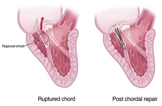
Over time, one of the flaps may rupture from its attachment to the heart muscle, leading to worse valve leakage. This type of mitral regurgitation is degenerative regurgitation.

Endocarditis

Another common cause of MR is endocarditis, a bacterial infection. It often occurs in people with pre-existing mitral valve prolapse.

Bacteria enter the bloodstream — often from dental procedures or poor oral health — and attach to the valve, causing an infection.

Taking antibiotics before you have dental work can help prevent infection.

Other well-known causes of mitral valve regurgitation

Other well-known causes of MR include:

  • Rheumatic fever. Can lead to scarring of the flaps and cause the valve to leak.
  • Heart attack or lack of blood supply to the heart muscle. This can stretch the heart's left lower chamber and the mitral valve, causing it to leak. This is functional or ischemic MR.
  • Irregular heart rhythms, such as AFib. These lead to stretching of the heart's left upper chamber and the mitral valve. This is also a type of functional or secondary MR.
  • Trauma. Can lead to a rupture of the valve apparatus.
  • Congenital malformation of the flaps. Some people are born with damage to the flaps, causing the valve to work improperly.
  • Radiation and certain drugs. Can damage the valve or affect how it works.
  • Connective tissue or systemic inflammatory diseases. Conditions such as Marfan syndrome, Ehlers-Danlos syndromes, scleroderma, and lupus have become linked to MR.

Mitral valve regurgitation risk factors

People with conditions that can harm the valve or flaps or affect the valve's function are at higher risk.

Factors that increase your risk of MR include:

  • Mitral valve prolapse or mitral stenosis.
  • Heart attack.
  • Certain medicines.
  • Age.
  • Rheumatic fever as a child.
  • Prior chest radiation.
  • Long-standing AFib.
  • Connective tissue disease, such as Ehlers-Danlos or Marfan syndrome.
  • Systemic inflammatory disease, such as scleroderma or lupus.

Mitral valve regurgitation complications

Without treatment, MR can lead to complications such as:

  • Heart failure requiring a hospital stay. MR causes the heart to work harder to pump blood throughout the body. Over time, the heart muscle weakens, causing it to fail.
  • Heart enlargement. The upper or lower left chamber can get larger or widen when the heart has to strain to pump blood.
  • Common heart rhythm problems such as AFib. Changes in heart rhythm can occur when mitral regurgitation causes the upper left chamber to get larger.
  • Rare heart rhythm problems such as premature ventricular contractions. Heart enlargement can lead to changes in the heart's rhythm.

Very rarely, MR caused by severe mitral valve prolapse can lead to sudden death.

Why choose UPMC's Center for Heart Valve Disease for mitral regurgitation care?

At the Center for Heart Valve Disease, we provide a team-based approach to:

  • Tailor mitral valve regurgitation treatment plans to each person's needs.
  • Help you enjoy an improved quality of life.
  • Provide the latest minimally invasive mitral valve techniques, such as the MitraClip®, backed by our own research.

Mitral Valve Regurgitation Symptoms and Diagnosis

Mitral valve regurgitation symptoms

MR can exist for years without causing major symptoms.

When symptoms do occur, they can include:

  • Shortness of breath, often worse with activity.
  • Fatigue or feeling tired.
  • Coughing — often a dry cough.
  • Swelling of feet or ankles.
  • Heart palpitations or feeling your own heartbeat.
  • Chest pain.
  • Anxiety.

When to see a doctor about mitral valve regurgitation

You should call a doctor if you have:

  • Trouble breathing when exercising, with exertion, or at rest.
  • New swelling of the legs, feet, or ankles.

Diagnosing mitral valve regurgitation

Your doctor at the UPMC Center for Heart Valve Disease will ask about your medical history and do a physical exam.

MR often causes murmurs or strange sounds of blood flowing through your heart.

Tests your doctor may use to diagnose MR include:

  • Echocardiogram. A heart ultrasound that uses sound waves to make images of the structures of your heart. This is often the first step.
  • Transesophageal echo (TEE). An outpatient test that uses sound waves like a standard echo but done within your esophagus. TEE gives doctors precise images of your heart valve, often in 3D. This is one of the best ways to diagnose MR correctly.
  • EKG. Measures the electrical activity of your heart.
  • Chest x-ray. Lets your doctor see the size and shape of your heart.
  • Heart catheterization. An outpatient test that lets the doctor see blood flow through the heart and its arteries.
  • MRI scan. Uses a magnet and radio waves to take pictures of your mitral valve.

Mitral Valve Regurgitation Treatment

Treatment for MR aims to limit or stop your valve leakage. It also relieves symptoms and improves the quality and length of your life.

For mild MR, medicine and close follow-up with frequent echocardiograms are often all you need.

But because MR is a mechanical problem with blood flow, valve repair or replacement is often best.

Doctors at the UPMC Center for Heart Valve Disease are experts in minimally invasive and surgical techniques for repairing your mitral valve.

Your doctor will use the approach that best meets your needs.
Mitral valve regurgitation occurs when the valve separating the left heart chambers doesn't close properly and allows blood to leak backward through the mitral valve. Treatment involves surgically repairing or replacing the mitral valve.

Valve repair surgery for mitral regurgitation

The first line treatment of MR, especially for mitral valve prolapse, is valve repair surgery.

This involves surgery to restore your mitral valve's normal function by rebuilding your existing flaps. We tailor the technique to your anatomy.

At the UPMC Center for Heart Valve Disease, we do this operation every day with a repair rate of over 90%.

Benefits of minimally invasive mitral valve surgery

Minimally invasive approaches make a small incision on the front of the chest or the right side of the chest.

These smaller cuts to access the heart lead to:

  • Less pain and blood loss.
  • Reduced risk of infection.
  • Faster recovery, allowing you to get back to your life sooner.

After mitral valve repair surgery, most people:

  • Get off the breathing machine quickly, sometimes while still in the OR.
  • Stay in the hospital for three to five days before going home.
  • Can do some activities the first three to four weeks post-op. But avoid heavy lifting and driving. Your doctor will explain what you can and can't do.

After three to four weeks of recovery, you can slowly return to normal healthy activity, including driving.

Minimally invasive robotic-assisted mitral valve surgery

UPMC is a leader in minimally invasive robotic mitral valve repair.

This allows the surgical team to access the mitral valve directly — often through a 2-inch incision — with technical and imaging precision.

It's the preferred treatment for people with isolated MR due to mitral valve prolapse.

Compared to some other types of surgery, robotic mitral valve repair:

  • Avoids disturbance to the ribs or chest.
  • Causes less pain.
  • Allows for a faster recovery and return to your daily routine.

Transcatheter Edge-to-Edge Repair (TEER)

UPMC is one of the few centers in southwestern Pa. to offer transcatheter edge-to-edge repair, a minimally invasive approach to MR valve repair.

Experts at UPMC's Center for Heart Valve Disease perform this procedure through a vein in the leg. The surgeon passes a catheter with the clip into the heart to bring the mitral valve's two flaps together.

UPMC uses two commercially available systems: Abbott's MitraClip System and Edwards' Pascal Precision System. Surgeons often use the TEER technique to treat people who are too high risk for heart valve surgery.

Mitral valve replacement surgery

Mitral repair is always our first approach, especially for those with degenerative or "floppy" valves.

But we may perform a mitral valve replacement for moderate to severe cases of MR.

This occurs if repairing the valve isn't an option because of:

  • Heavy calcium.
  • Infection.
  • Severe stretching from heart failure.

We replace the mitral valve with a valve made of either metal flaps (mechanical) or cow or pig tissue (biologic).

  • Metal valves are long-lasting but require you to take a blood thinner, warfarin (Coumadin®), for life.
  • Tissue valves don't last as long as metal ones but often don't need lifelong blood thinners. Depending on your age and condition at the time of surgery, tissue valves may eventually need replacement.