![Brain fiber image](https://dam.upmc.com/-/media/upmc/media/media-kit/hdft/publishingimages/thumbnails/brain.png?rev=c6b3eca91c9c4782a443b985bcdf27ac&hash=BF3AD3B74C106084E89E4993D1C06872) |
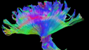
High definition fiber-tracking of a million brain fibers.
Credit: Walt Schneider Laboratory |
![Loss of fibers image](https://dam.upmc.com/-/media/upmc/media/media-kit/hdft/publishingimages/thumbnails/tbi-damage-midbrain-break.png?rev=1f7821c3879f48be91bdc215524bb004&hash=DF31E2036190DAC4BE4C19095BEBA41F) |
High definition fiber tracking reveals loss of fibers, or connections, on the injured right side (yellow) and the intact, undamaged left side (green). The patient was injured in an ATV accident and lost function in his left leg, arm and hand.
Credit: Walt Schneider Laboratory |
![Brain fibers image](https://dam.upmc.com/-/media/upmc/media/media-kit/hdft/publishingimages/thumbnails/tbi-damage-view-roated-see-left-difference.png?rev=a996a676c14e46fea44726fd66b75370&hash=B95EFE0DAF996227A068BA40922743A0) |
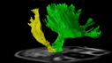
This shows the brain fibers that travel from the cortex to the lower brain, including fibers that control movement of leg, arm and hand. The uninjured side of the patient’s brain is in green. The injured side is in yellow and shows the fiber loss that has led to the patient’s difficulty moving his left arm and hand.
Credit: Walt Schneider Laboratory |
![Brain fibers imaging](https://dam.upmc.com/-/media/upmc/media/media-kit/hdft/publishingimages/thumbnails/tbi-uninjured-left-side-caronaradiata2-rgb.png?rev=6e5fc469dd854773ab32048541c49fb7&hash=E03C562BB5A81D789AAA60B0411DA26D) |
Brain fibers that control right-sided limb movement were not damaged when the patient was hurt in an ATV accident.
Credit: Walt Schneider Laboratory |
![Brain fibers image](https://dam.upmc.com/-/media/upmc/media/media-kit/hdft/publishingimages/thumbnails/tbi-injured-right-rgb-caronaradiata.png?rev=c5c1870b3a43425abb129949700ab276&hash=6B8685F62CB4AF23FC76CC955841D060) |
The patient injured in an ATV accident lost brain fibers, or connections, that control his left leg, arm and hand, leading to their impaired movement.
Credit: Walt Schneider Laboratory |
![Doctor Schneider](https://dam.upmc.com/-/media/upmc/media/media-kit/hdft/publishingimages/thumbnails/waltschneider.png?rev=22ce07a35c7341ebbc1edb6495b1931f&hash=3971160C16AB7CA6810D10BC482F8291) |
Walter Schneider, Ph.D., professor of psychology, University of Pittsburgh Learning Research and Development Center. |
![Doctor Okonkwo with a patient](https://dam.upmc.com/-/media/upmc/media/media-kit/hdft/publishingimages/thumbnails/okonkwo-checks-daniel.png?rev=3f518fe59ce84bcfb0846cb88cfbb1a9&hash=B83363668712F50CDE04BBAA7DF16D6D) |
David Okonkwo, M.D., associate professor, Department of Neurological Surgery, Pitt School of Medicine, examines Daniel Stunkard, who sustained a traumatic brain injury in an ATV accident. |
![Doctor Boada](https://dam.upmc.com/-/media/upmc/media/media-kit/hdft/publishingimages/thumbnails/walt-fernando-viewmri.png?rev=b5a1a20f6e304499997ce3bde578906f&hash=E9E4ABAF67E8835334D93172BD323756) |
Fernando Boada, M.D., associate professor of radiology and bioengineering, and director of the MR Research Center, University of Pittsburgh, and Walter Schneider, Ph.D., professor of psychology, Pitt’s Learning Research and Development Center. |
![Daniel Stunkard lifting his arm](https://dam.upmc.com/-/media/upmc/media/media-kit/hdft/publishingimages/thumbnails/daniel-moves-arm.png?rev=499b866f3da149eb9a982fa6b76a8d82&hash=26CDEA20D41E2ED784B395D630362052) |
Daniel Stunkard demonstrates his difficulty in lifting his left arm. He sustained a traumatic brain injury in an ATV accident. |
![Daniel Stunkard using his left arm](https://dam.upmc.com/-/media/upmc/media/media-kit/hdft/publishingimages/thumbnails/daniel-pegboardcrop.png?rev=8ad88145a6154cfaa5ba88f2b33cec79&hash=B3D659B2D278D20FC0B165C362CA1BFB) |
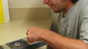
Daniel Stunkard attempts to place small pegs in a board with his left hand, which is impaired due to a traumatic brain injury. |